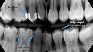
Dental health plays a pivotal role in our overall well-being. One of the most common dental issues faced by people worldwide is cavities, also known as dental caries. Cavities are small holes or decayed areas in the hard surface of teeth caused by prolonged exposure to acids produced by bacteria in the mouth. While they can often be detected during a routine dental exam, dental X-rays provide a more in-depth view, allowing dentists to identify cavities that may not be visible to the naked eye. But what does a cavity look like on an X-ray? Let’s dive into the details to understand this better.
Understanding Dental X-Rays
Dental X-rays, or radiographs, are diagnostic tools that help dentists visualize the interior of teeth, the roots, surrounding bone, and the position of teeth within the jaw. There are several types of dental X-rays, but the two most commonly used for detecting cavities are bitewing X-rays and periapical X-rays:
- Bitewing X-Rays: These are taken with the patient biting down on a wing-shaped device. Bitewing X-rays focus on the crowns of the upper and lower teeth, making them ideal for detecting cavities between teeth.
- Periapical X-Rays: These provide a detailed view of the entire tooth, from crown to root, and the surrounding bone structure. They are often used to check for issues below the gumline.
Both types of X-rays use minimal radiation and are considered safe for most patients. The images captured provide a black, white, and gray visual representation of the teeth and surrounding structures.
How Cavities Appear on X-Rays
On an X-ray, a healthy tooth appears mostly white because its dense enamel and dentin absorb X-rays effectively. The surrounding tissues and gums typically appear darker. When a cavity forms, the decayed area becomes less dense than healthy tooth structures, allowing more X-rays to pass through. This results in a darker spot or shadow on the X-ray image.
Key Features of Cavities on X-Rays:
- Darkened Areas: Cavities typically show up as dark spots or shadows on the lighter image of the tooth. The size and shape of the shadow depend on the extent of the decay.
- Location:
- Interproximal Cavities: Cavities that form between teeth are easier to spot on bitewing X-rays. These appear as small, dark, triangular shapes near the contact point between teeth.
- Occlusal Cavities: Cavities on the biting surface may not always be visible on X-rays unless they are deep. When visible, they appear as dark spots in the grooves of the tooth.
- Root Cavities: Decay on the roots, often seen in older adults or those with gum recession, appears as a darkened area near the root surface.
- Depth of Decay: The darkness and size of the shadow can indicate the severity of the cavity. Shallow cavities might appear as faint shadows, while deeper ones create more pronounced dark areas.
- Margins: The edges of a cavity on an X-ray may appear irregular or fuzzy, contrasting with the smooth appearance of healthy enamel.
Differentiating Cavities from Other Issues
Not every dark spot on an X-ray is a cavity. Dentists are trained to distinguish between cavities and other conditions that may look similar, such as:
- Tartar (Calculus): Hardened plaque can sometimes appear dark on X-rays but has a different shape and texture than decay.
- Restorations: Fillings, crowns, and other dental work may cast shadows, which can resemble cavities.
- Wear and Tear: Attrition or erosion may mimic decay but often has a different pattern on X-rays.
- Defects or Anomalies: Natural pits, grooves, or developmental defects in teeth may also appear dark.
To make an accurate diagnosis, dentists combine X-ray findings with a clinical examination and the patient’s dental history.
Importance of Early Detection
Detecting cavities early is crucial to preventing further damage to the tooth. Left untreated, cavities can progress to involve the deeper layers of the tooth, leading to:
- Pain and Sensitivity: As decay reaches the dentin and pulp, it can cause discomfort.
- Infection: Severe decay can lead to abscesses or infections, which may require root canal treatment or extraction.
- Structural Damage: Advanced cavities can weaken the tooth, increasing the risk of fractures.
X-rays play a vital role in early detection, especially for cavities in hard-to-see areas like between teeth or below existing fillings.
What Happens After a Cavity is Detected?
Once a cavity is identified on an X-ray, the dentist will recommend a course of action based on its size and severity:
- Small Cavities: These may be treated with fluoride treatments or dental sealants to prevent further decay.
- Moderate Cavities: Fillings are the most common treatment for cavities that have progressed beyond the enamel.
- Severe Cavities: When decay reaches the pulp, a root canal may be necessary. In extreme cases, the tooth may need to be extracted and replaced with a dental implant or bridge.
Maintaining Good Dental Health
Preventing cavities begins with good oral hygiene practices and regular dental check-ups. Here are some tips to maintain healthy teeth and reduce the need for X-rays to diagnose cavities:
- Brush and Floss Regularly: Brush twice a day with fluoride toothpaste and floss daily to remove food particles and plaque.
- Limit Sugary Foods and Drinks: Reduce consumption of sweets and sugary beverages that feed harmful bacteria in the mouth.
- Use Fluoride: Fluoride strengthens enamel and helps prevent cavities.
- Visit Your Dentist Regularly: Routine dental visits ensure early detection of any issues.
- Consider Dental Sealants: These protective coatings can shield teeth from decay, especially in children and teens.
Conclusion
Dental X-rays are indispensable tools in modern dentistry, providing a clear view of hidden cavities and other dental issues. A cavity on an X-ray typically appears as a darkened area contrasting with the lighter, denser structure of a healthy tooth. Recognizing and treating cavities early can save time, money, and discomfort while preserving your natural teeth.
If you suspect a cavity or it’s time for a routine dental check-up, don’t hesitate to schedule an appointment with your dentist. Regular care and vigilance are the keys to a bright, healthy smile.
